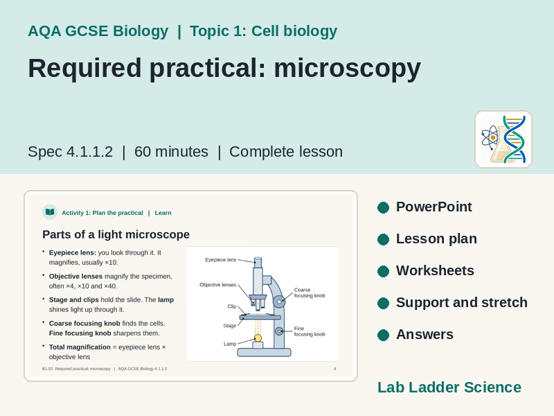
Microscopy required practical lesson for AQA GCSE Biology 4.1.1.2. Prepare slides, observe, draw and estimate cell size. PowerPoint, worksheets, answers.
What is this resource?
This is a complete lesson on the microscopy required practical for AQA GCSE Biology (8461), specification point 4.1.1.2 (Required practical activity 1). It is the second lesson in Topic 1: Cell biology and is designed to last 60 minutes.
Students learn the parts of a light microscope, calculate total magnification, prepare a slide of onion epidermis cells, observe plant and animal cells at different magnifications, and make labelled scientific drawings with a magnification. They then use the field of view to estimate the real size of a cell and answer exam-style analysis questions.
What’s included
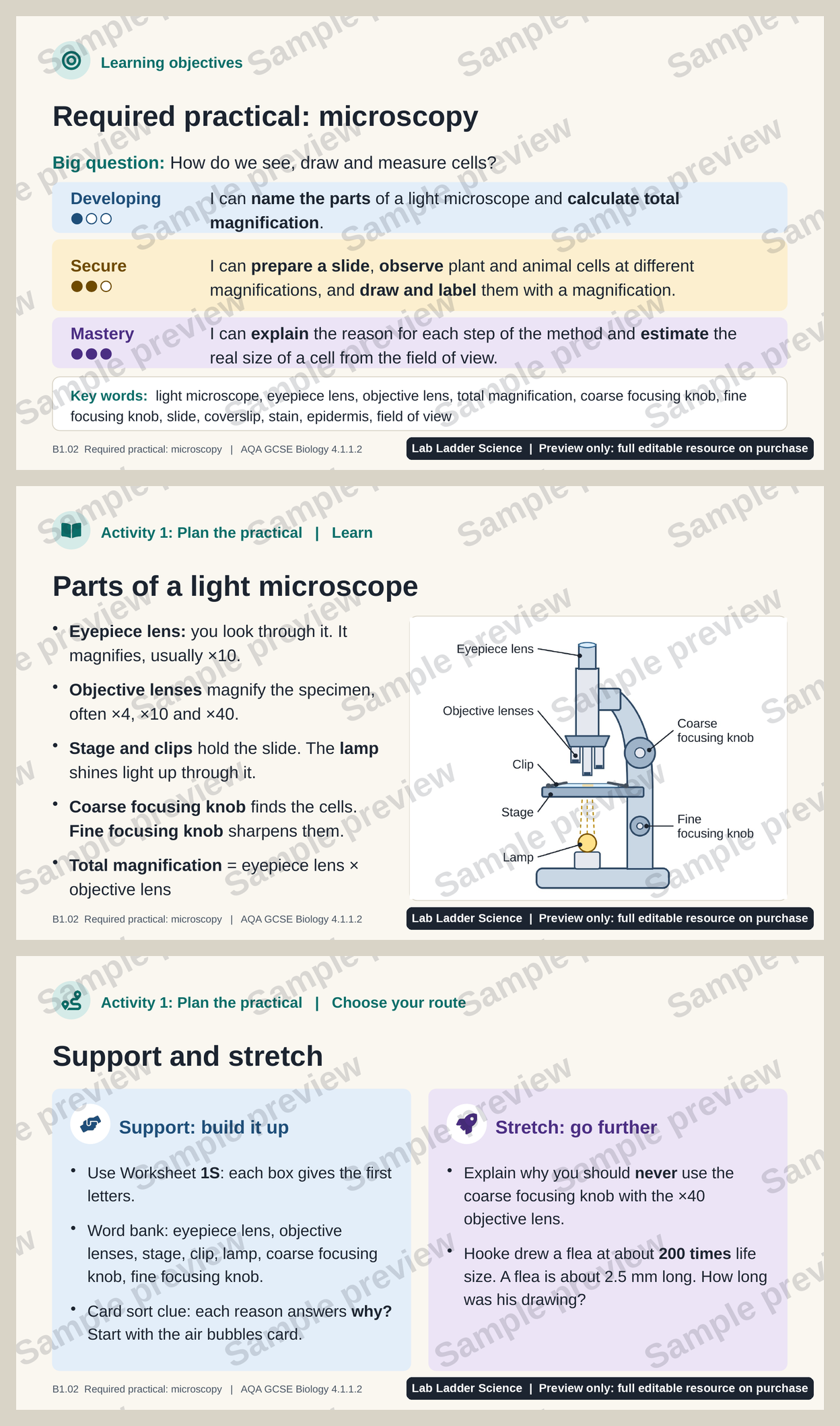
• PowerPoint lesson (33 slides) with full teacher notes on every slide: timings, the full method, technician list, safety, answers, misconceptions and questioning prompts.
• Starter on screen as students arrive, based on a real-world hook: Robert Hooke’s first drawings of cells in 1665.
• Learning objectives at three levels of challenge: Developing, Secure and Mastery.
• Three learning activities, each with a core task, a support option, a stretch task and a ‘stand back’ mini-whiteboard check with answers.
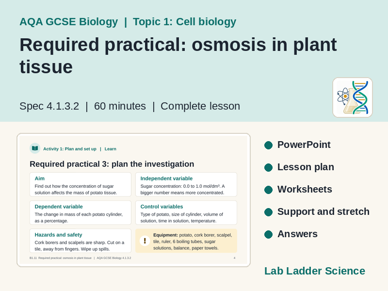
• Required practical detail: aim, equipment, method, independent, dependent and control variables, hazards and precautions, and a results table.
• Clear original diagrams: a labelled light microscope, slide preparation steps, a field of view next to a model scientific drawing, and a worked example of estimating cell size.
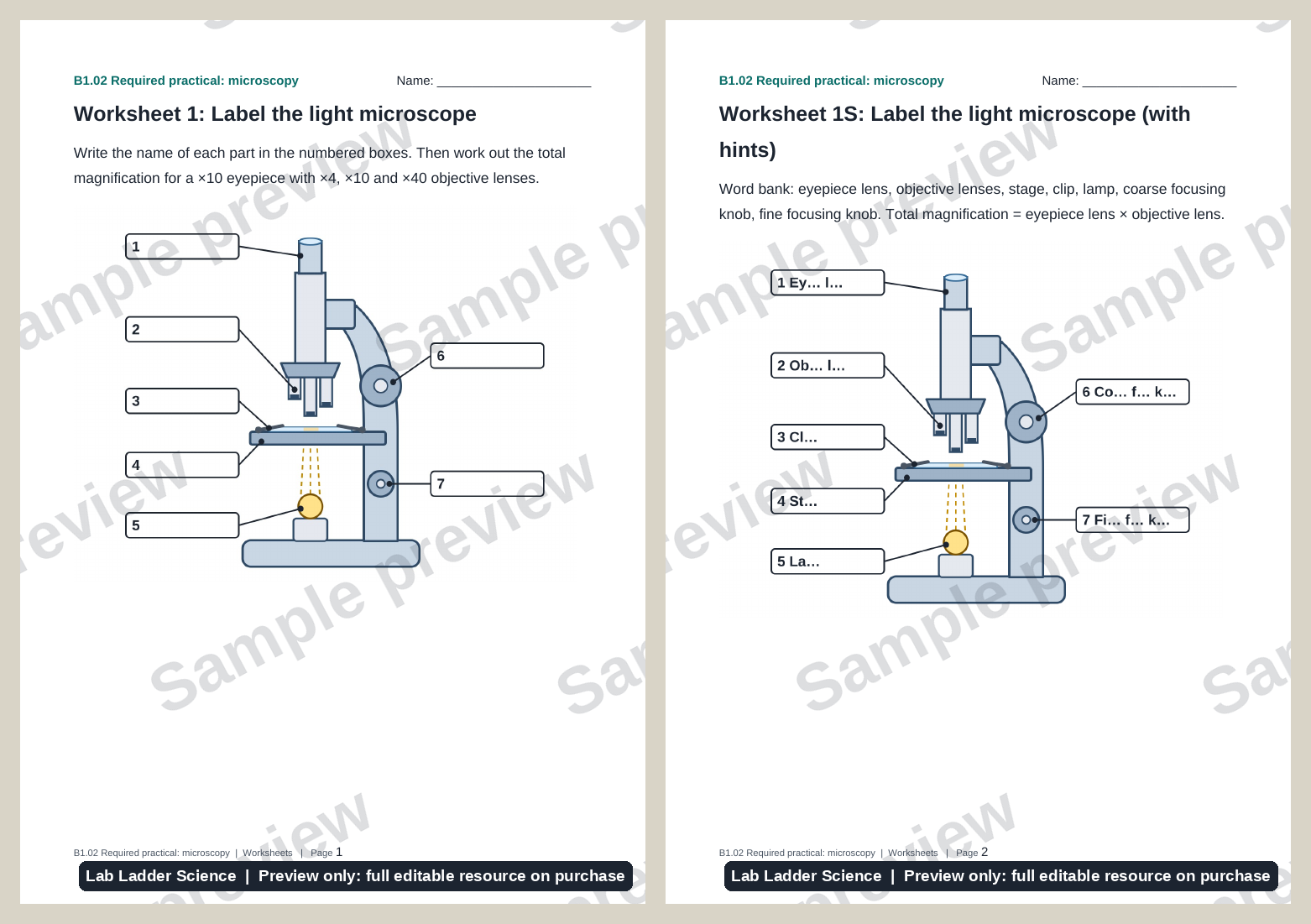
• Editable Word worksheets: label the microscope (core and support versions), a method steps and reasons card sort, a results table, and exam-style analysis questions (core and support versions).
• Plenary exit ticket with one question per level, an answers slide for self-assessment and a return to the starter.
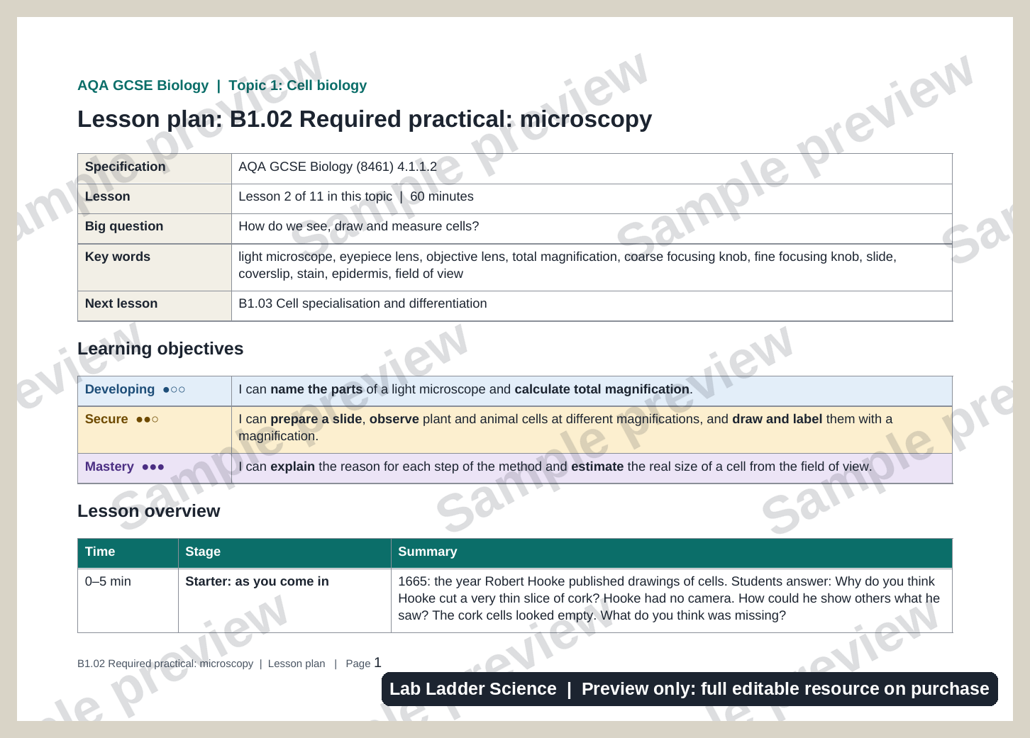
• Word lesson plan with objectives, lesson sequence, differentiation, answers and common misconceptions.
Lesson outline
• Starter (5 min): Why did Robert Hooke cut such a thin slice of cork, and why did his cells look empty?
• Activity 1 (12 min): Label a light microscope, calculate total magnification and match each method step to its reason.
• Activity 2 (20 min): Prepare a slide of onion epidermis, observe onion and animal cells at ×40, ×100 and ×400, record results and draw the cells.
• Activity 3 (13 min): Estimate cell size from the field of view and answer exam-style questions on a set of results.
• Plenary (8 min): Exit ticket at three levels, then self-assessment against the objectives.
Differentiation and inclusion
Every activity offers support and stretch that all students can choose, so no one is singled out. Support includes first-letter hints, a word bank, a drawing checklist and a step-by-step version of the analysis questions. Stretch tasks ask students to add a scale bar, explain why the coarse focusing knob is never used at high power, tell cells from air bubbles and evaluate the accuracy of their estimate.
Taking a cheek cell sample is always optional, with prepared slides as an equal alternative. The slides follow the British Dyslexia Association Style Guide: a clear sans serif font at a large size, a soft cream background, bold (not italics) for emphasis, left-aligned text and plain English. Levels of challenge never rely on colour alone, every slide has a proper title for screen readers, and diagrams include alt text.
How you could use it
• Teach it as the required practical lesson straight after a lesson on animal and plant cells.
• Use the card sort and analysis questions to revise the practical before mocks, even without microscopes.
• Plan the practical with your technician: the notes include the full method, equipment list and safety points.
• Set the exam-style questions on Worksheet 4 as homework.
Specification coverage
AQA GCSE Biology 8461, section 4.1.1.2 Animal and plant cells, Required practical activity 1: use a light microscope to observe, draw and label a selection of plant and animal cells, including a magnification scale. Key words: light microscope, eyepiece lens, objective lens, total magnification, focusing, slide, coverslip, stain, epidermis, field of view, scientific drawing. The previous lesson covers animal and plant cell structure, and the next lesson covers cell specialisation and differentiation.
You must log in and be a buyer of this download to submit a review.